Add new comment

Warning message

All forms are disabled.

Immunocytochemistry on primary cultures

fluorecent image of cultured neurons and glial cells

Group meeting brief: June 24, 2014

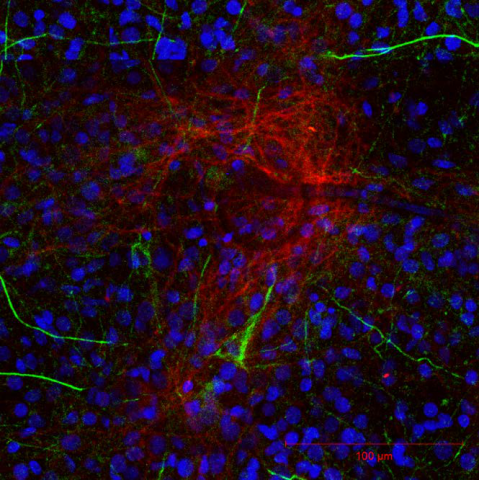
In his talk José showcased he and Webber's progress on transfecting the neurons with fluorescent proteins and imaging them with confocal microscopy. The dendrites and axons of neurons are transfected with microtubule-associated protein 2 and neuronfilament 200 which glow in red and green respectively. While, the nuclei of the cells fluoresce in blue with DAPI. The images are clear and can possibly give information on connectivity and correlate with electrophysiological results with future improvement. Some problems in the penetrating the bulk of cell cluster likely can be improve by reducing the planting density.

* Login to see slides.


Filtered HTML

  • Web page addresses and e-mail addresses turn into links automatically.
  • Allowed HTML tags: <a> <em> <strong> <cite> <blockquote> <code> <ul> <ol> <li> <dl> <dt> <dd>
  • Lines and paragraphs break automatically.
  • Mathematics inside the configured delimiters is rendered by MathJax. The default math delimiters are $$...$$ and \[...\] for displayed mathematics, and $...$ and \(...\) for in-line mathematics.

Plain text

  • No HTML tags allowed.
  • Web page addresses and e-mail addresses turn into links automatically.
  • Lines and paragraphs break automatically.
CAPTCHA
This question is for testing whether or not you are a human visitor and to prevent automated spam submissions.
Image CAPTCHA
Enter the characters shown in the image.